M.D. Smoller - Inflammatory Dermatoses: The Basics
Here you can read online M.D. Smoller - Inflammatory Dermatoses: The Basics full text of the book (entire story) in english for free. Download pdf and epub, get meaning, cover and reviews about this ebook. year: 2010, publisher: Springer Nature, genre: Detective and thriller. Description of the work, (preface) as well as reviews are available. Best literature library LitArk.com created for fans of good reading and offers a wide selection of genres:
Romance novel
Science fiction
Adventure
Detective
Science
History
Home and family
Prose
Art
Politics
Computer
Non-fiction
Religion
Business
Children
Humor
Choose a favorite category and find really read worthwhile books. Enjoy immersion in the world of imagination, feel the emotions of the characters or learn something new for yourself, make an fascinating discovery.
- Book:Inflammatory Dermatoses: The Basics
- Author:
- Publisher:Springer Nature
- Genre:
- Year:2010
- Rating:3 / 5
- Favourites:Add to favourites
- Your mark:
Inflammatory Dermatoses: The Basics: summary, description and annotation
We offer to read an annotation, description, summary or preface (depends on what the author of the book "Inflammatory Dermatoses: The Basics" wrote himself). If you haven't found the necessary information about the book — write in the comments, we will try to find it.
M.D. Smoller: author's other books
Who wrote Inflammatory Dermatoses: The Basics? Find out the surname, the name of the author of the book and a list of all author's works by series.


Inflammatory Dermatoses: The Basics — read online for free the complete book (whole text) full work
Below is the text of the book, divided by pages. System saving the place of the last page read, allows you to conveniently read the book "Inflammatory Dermatoses: The Basics" online for free, without having to search again every time where you left off. Put a bookmark, and you can go to the page where you finished reading at any time.
Font size:
Interval:
Bookmark:
- Superficial perivascular dermatitis
- Inflammatory dermatoses involving venules in superficial vascular plexus
- Other histologic changes help with further classification
- Superficial perivascular dermatitis (SPD)
- Without epidermal changes
- Lymphocytic infiltrate
- Mixed infiltrate
- With epidermal changes
- Interface/vacuolar and lichenoid dermatitis ()
- Spongiotic dermatitis ()
- Psoriasiform dermatitis ()Table 1.1Superficial perivascular lymphohistiocytic dermatitis without epidermal changesPigmented purpuric eruptionViral exanthemGyrate erythemaDermatophytosesPost-inflammatory pigment alterationRocky mountain spotted feverPolymorphous light eruption
- Pigmented purpuric eruption, Schamberg variant (progressive pigmentary dermatosis)
- Clinical
- Erythematous, non-blanching patches
- Usually on lower extremities, pre-tibial
- Most common in middle-aged men
- May be related to drug exposure in some cases
- Controversial relationship with mycosis fungoides
- Recent literature suggests possibility of progression
- Multiple subtypes of pigmented purpuric eruption
- This is the most common; all other subtypes demonstrate epidermal changes
- Histologic findings
- Superficial perivascular lymphohistiocytic infiltrate
- Eosinophils not common
- Mild spongiosis and exocytosis
- Hemorrhage and hemosiderin surrounding vessels in superficial vascular plexus
- Perls iron or Prussian blue stain often helpful in demonstrating dermal hemosiderin deposition (necessary to document chronicity of process) (Figs. )
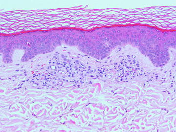 Fig. 1.1Pigmented purpuric eruption, Schamberg variant, shows a mild superficial perivascular lymphohistiocytic infiltrate. Erythrocyte extravasation is present. The overlying epidermis is uninvolved
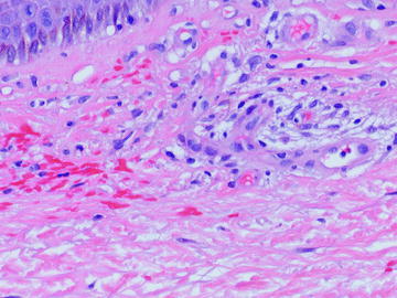 Fig. 1.2Pigmented purpuric eruption, Schamberg variant. This high-power image shows perivascular erythrocyte extravasation. Hemosiderosis is variable, depending on the duration of disease, and can be nearly non-existent as in this case
- Viral exanthem
- Clinical
- Morbilliform (measles-like) eruption
- Erythematous papules and macules usually rapid onset
- Resolves rapidly without sequelae in most cases
- Histologic findings
- Superficial perivascular lymphohistiocytic infiltrate
- Inflammation does not usually extend into deeper dermis
- Eosinophils very uncommon
- Slight exocytosis, epidermal spongiosis, and basal vacuolopathy
- Occasional dying keratinocytes, but very few
- Non-specific findings hard to establish diagnosis without clinical correlation (Figs. )
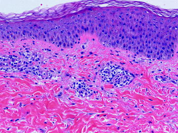 Fig. 1.3This viral exanthem shows a superficial perivascular lymphohistiocytic infiltrate with no alterations in the overlying epidermis
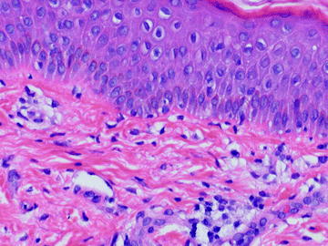 Fig. 1.4Mild spongiosis and interface degeneration are seen in this viral exanthem
- Gyrate erythema
- Clinical
- Most commonly refers to erythema annulare centrifugum, but also includes erythema gyratum repens, erythema chronicum migrans, other less common eruptions
- Annular, erythematous lesions on trunk
- Slow outward extension of plaques in some cases
- Peripheral, delicate scale
- Histologic findings
- Almost entirely lymphoid infiltrate in a perivascular distribution
- Eosinophils may rarely present in small numbers
- Tight cuffing of lymphocytes around vessels of the superficial vascular plexus
- Some cases also involve deeper vascular plexus
- Scant parakeratotic scale with mild underlying spongiosis if peripheral scale is biopsied
- Plasma cells present in small numbers in erythema chronicum migrans, but not usually in erythema annulare centrifugum (Figs. )
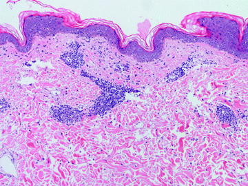 Fig. 1.5Erythema annulare centrifugum is characterized by a lymphohistiocytic infiltrate tightly cuffed around the vessels
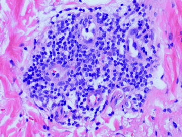 Fig. 1.6Erythema annulare centrifugum characteristically has a lymphohistiocytic infiltrate. Plasma cells may be seen; neutrophils and eosinophils are not characteristic
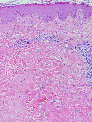 Fig. 1.7Erythema chronicum migrans shows a superficial perivascular lymphohistiocytic infiltrate without significant epidermal involvement
- Dermatophytosis
- Clinical
- Tinea versicolor, caused by Pityrosporum versicolor , classically shows minimal epidermal change
Font size:
Interval:
Bookmark:
Similar books «Inflammatory Dermatoses: The Basics»
Look at similar books to Inflammatory Dermatoses: The Basics. We have selected literature similar in name and meaning in the hope of providing readers with more options to find new, interesting, not yet read works.
Discussion, reviews of the book Inflammatory Dermatoses: The Basics and just readers' own opinions. Leave your comments, write what you think about the work, its meaning or the main characters. Specify what exactly you liked and what you didn't like, and why you think so.