Bruce R. Smoller - Dermal Tumors: The Basics
Here you can read online Bruce R. Smoller - Dermal Tumors: The Basics full text of the book (entire story) in english for free. Download pdf and epub, get meaning, cover and reviews about this ebook. year: 2011, publisher: Springer Verlag, genre: Detective and thriller. Description of the work, (preface) as well as reviews are available. Best literature library LitArk.com created for fans of good reading and offers a wide selection of genres:
Romance novel
Science fiction
Adventure
Detective
Science
History
Home and family
Prose
Art
Politics
Computer
Non-fiction
Religion
Business
Children
Humor
Choose a favorite category and find really read worthwhile books. Enjoy immersion in the world of imagination, feel the emotions of the characters or learn something new for yourself, make an fascinating discovery.
- Book:Dermal Tumors: The Basics
- Author:
- Publisher:Springer Verlag
- Genre:
- Year:2011
- Rating:5 / 5
- Favourites:Add to favourites
- Your mark:
Dermal Tumors: The Basics: summary, description and annotation
We offer to read an annotation, description, summary or preface (depends on what the author of the book "Dermal Tumors: The Basics" wrote himself). If you haven't found the necessary information about the book — write in the comments, we will try to find it.
Dermal Tumors: The Basics will serve as an effective and efficient handbook for the student of dermatopathology, and as a practical bench reference for the practicing diagnostician who desires rapid access to criteria that are useful in differentiating histologically similar entities. The reader will be able to focus upon a single histologic observation, i.e., inflammatory conditions without epidermal changes, and use this as a starting point from which to build a differential diagnosis based upon pattern recognition. As each entity is addressed, there will be a concise discussion of the basic clinical findings and epidemiologic associations. This will be followed by a histologic description, highlighting areas that serve to discriminate between the entity under discussion and similar ones. Any immunologic studies that might augment the diagnostic sensitivity or specificity will be discussed.
The chapters are thematically based and consist of essential bullet points arranged in organized outlines allowing for easy access and direct comparison between entities. The salient histologic features are depicted with abundant high quality, full-color photomicrographs placed immediately adjacent to the appropriate histologic bullet points. This volume will serve as an effective and efficient handbook for the student of dermatopathology, and as a practical bench reference for the practicing diagnostician who desires rapid access to criteria that are useful in differentiating histologically similar entities. The elaborate pictorial documentation will also enable the book to serve as an atlas of the commonest dermatologic disorders.
Bruce R. Smoller: author's other books
Who wrote Dermal Tumors: The Basics? Find out the surname, the name of the author of the book and a list of all author's works by series.


Dermal Tumors: The Basics — read online for free the complete book (whole text) full work
Below is the text of the book, divided by pages. System saving the place of the last page read, allows you to conveniently read the book "Dermal Tumors: The Basics" online for free, without having to search again every time where you left off. Put a bookmark, and you can go to the page where you finished reading at any time.
Font size:
Interval:
Bookmark:
- Classifications of cutaneous lymphomas
- Two recent systems both had problems
- Revised European-American Classification of Lymphomas (REAL)
- European Organization for Research and Treatment of Cancer (EORTC)
- REAL classification
- Uses classification of systemic lymphomas and applies them to cutaneous lymphomas
- Classification based upon pathologic, genetic, and clinical features and adds immunohistochemical criteria
- Cutaneous involvement accounted for in clinical features
- EORTC classification
- Recognized cutaneous lymphomas as a distinct entity
- Organ-based classification system
- Only system that has been clinically validated for this group of diseases; classified as indolent, intermediate or, aggressive
- WHO-EORTC classification
- Consensus conference in 2005 incorporated features of both systems to acknowledge particulars of cutaneous lymphomas
- WHO 2008 classification
- Integrated the WHO-EORTC classification of cutaneous lymphomas in the general classification
- Only minor changes in terminology, except blastic plasmacytoid dendritic cell neoplasm, previously CD4+/CD56+ hematodermic neoplasm, previously blastic NK cell lymphoma
- Overview
- 3% per year increase in incidence of cutaneous lymphomas every year since 1970
- Cutaneous lymphomas represent a very heterogeneous group of diseases with very different clinical presentations, courses, treatment programs, and prognoses
- Second most common (after GI tract) site of extra-nodal non-Hodgkins lymphomas
- Large groupings include T cell lymphomas, B cell lymphomas, and Hodgkins lymphoma
- Overall incidence estimate of 1:100,000
- 75% of cutaneous lymphomas are T cell lymphomas, 25% B cell lymphomas
- Hodgkins lymphoma is exceedingly rare in skin
- 510% of T cell lymphomas are not mycosis fungoides
- T cell lymphomas (Table )Table 1.1T cell lymphomasMycosis fungoides/Sezary syndromePrimary cutaneous CD30+ lymphoproliferative disordersSubcutaneous panniculitis-like T cell lymphomaExtranodal NK/T cell lymphoma, nasal typeBlastic plasmacytoid dendritic cell neoplasm(Peripheral T cell lymphoma)
- Mycosis fungoides (MF)/Sezary syndrome
- Epidemiologic features
- 0.42 cases/100,000 population
- Overall, more than 50% of all cutaneous lymphomas
- Incidence rapidly increased in 1970s1980s, but now stabilized
- Twice as common in African Americans as in Caucasian Americans
- Twice as common in men as in women
- Associated with 3.3X relative risk for second, non-cutaneous malignancy
- Clinical features
- Usual onset in middle age (mean 5560), but can appear at any age
- Starts most commonly as erythematous, slightly scaly patches on flank and buttocks
- Progress to plaques and ultimately tumors (in a minority of patients)
- Poikilodermatous variant uncommon
- Involvement of lymph nodes, spleen, liver only late in course
- Histologic features (Figs. )
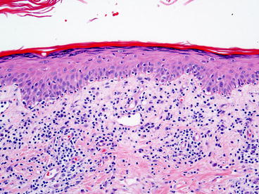 Fig. 1.1Early lesions of mycosis fungoides show an epidermotropic lymphocytic infiltrate. Single atypical lymphocytes are seen lining up along the basilar epidermis
 Fig. 1.2In more evolved lesions of mycosis fungoides, small clusters of atypical lymphocytes and increased single lymphocytes are present in the epidermis which also shows only minimal spongiosis and no dyskeratosis
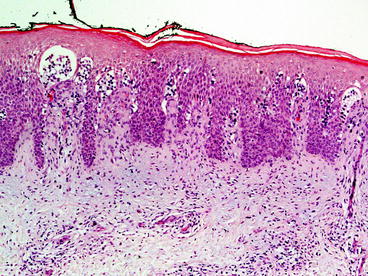 Fig. 1.3This case of mycosis fungoides shows only a mild dermal infiltrate but numerous Pautriers microabscesses
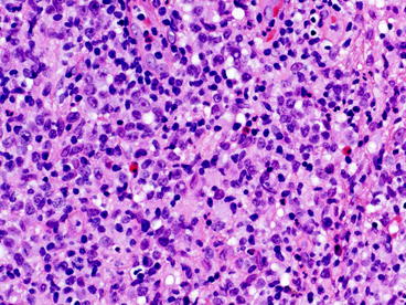 Fig. 1.4Variable number of eosinophils and some plasma cells are often present in the dermal infiltrate of mycosis fungoides
- Epidermotropic proliferation of hyerconvoluted, hyperchromatic lymphocytes in clusters (Pautriers microabscesses) and as single cells within epidermis
- Difficult diagnosis to make in early lesions
- Later lesions with less epidermotropism and more cytologic atypia
- Variable number of eosinophils and some plasma cells
- Immunologic features (Figs. )
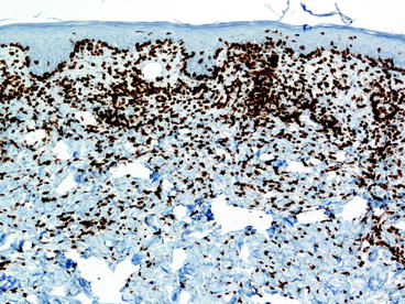 Fig. 1.5The epidermotropic lymphocytes in mycosis fungoides most commonly express CD4
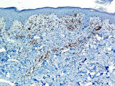 Fig. 1.6Loss of normal T-cell markers such as CD5 is common as in this case of mycosis fungoides
- Mycosis fungoides is defined as a neoplastic proliferation of CD3+, CD4+ T helper lymphocytes
- CD8+ tumors are rare but are now classified as MF
- Loss of CD2, CD3, and CD5 often seen
- Clonal rearrangements of T cell receptor is ultimately detected in most cases, but only 50% of early lesions.
- Molecular features
- T-cell gene rearrangements seen in 5080% of patch-stage lesions, nearly 100% of tumor stage and Sezary cases
Font size:
Interval:
Bookmark:
Similar books «Dermal Tumors: The Basics»
Look at similar books to Dermal Tumors: The Basics. We have selected literature similar in name and meaning in the hope of providing readers with more options to find new, interesting, not yet read works.
Discussion, reviews of the book Dermal Tumors: The Basics and just readers' own opinions. Leave your comments, write what you think about the work, its meaning or the main characters. Specify what exactly you liked and what you didn't like, and why you think so.