M.D. Smoller - Epidermal Cell Tumors: The Basics
Here you can read online M.D. Smoller - Epidermal Cell Tumors: The Basics full text of the book (entire story) in english for free. Download pdf and epub, get meaning, cover and reviews about this ebook. year: 2010, publisher: Springer Nature, genre: Detective and thriller. Description of the work, (preface) as well as reviews are available. Best literature library LitArk.com created for fans of good reading and offers a wide selection of genres:
Romance novel
Science fiction
Adventure
Detective
Science
History
Home and family
Prose
Art
Politics
Computer
Non-fiction
Religion
Business
Children
Humor
Choose a favorite category and find really read worthwhile books. Enjoy immersion in the world of imagination, feel the emotions of the characters or learn something new for yourself, make an fascinating discovery.

- Book:Epidermal Cell Tumors: The Basics
- Author:
- Publisher:Springer Nature
- Genre:
- Year:2010
- Rating:5 / 5
- Favourites:Add to favourites
- Your mark:
Epidermal Cell Tumors: The Basics: summary, description and annotation
We offer to read an annotation, description, summary or preface (depends on what the author of the book "Epidermal Cell Tumors: The Basics" wrote himself). If you haven't found the necessary information about the book — write in the comments, we will try to find it.
M.D. Smoller: author's other books
Who wrote Epidermal Cell Tumors: The Basics? Find out the surname, the name of the author of the book and a list of all author's works by series.


Epidermal Cell Tumors: The Basics — read online for free the complete book (whole text) full work
Below is the text of the book, divided by pages. System saving the place of the last page read, allows you to conveniently read the book "Epidermal Cell Tumors: The Basics" online for free, without having to search again every time where you left off. Put a bookmark, and you can go to the page where you finished reading at any time.
Font size:
Interval:
Bookmark:
- Benign melanocytic proliferations
- Also known as melanocytic nevi, moles
- Nevus means hamartoma and is likely a misnomer and nevi have been shown to be true clonal proliferations
- Present at birth, but most arise during adolescence or early adulthood
- Only rarely arise later in life (after age 40)
- Present in vast majority of Caucasians, also present in other racial groups
- Potential for malignant transformation is less than 1/100,000 in acquired melanocytic nevi
- Benign melanocytic proliferations (see Table )Table 1.1Benign melanocytic proliferationsCommon acquired melanocytic nevusCongenital melanocytic nevusHalo nevusNevus of special sites (acral, genital)Combined nevusBalloon cell nevusVolume III,Spindle and epithelioid cell (Spitz) nevusVolume III,Blue nevusVolume II, Chapter 12
- Common melanocytic nevus
- Proposed life cycle for melanocytic proliferations (including common acquired, congenital, dysplastic or atypical, Spitz, acral)
- Clinical
- Junctional nevus flat, deeply pigmented lesions with sharp edges, usually oval or circular
- Compound nevus raised above surface of skin, retain pigmentation
- Intradermal nevus nodular to polypoid, lose pigment (skin-colored)
- Histologic
- Junctional nevus proliferation of melanocytes confined to the epidermis, largely nested along basement membrane (Figs. )
- Compound nevus some melanocytes drop into dermis and some remain in the epidermis (Fig. )
- Intradermal nevus intraepidermal component of melanocytic proliferation is absent; all residual melanocytes are within dermis (Fig. )
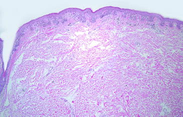 Fig. 1.1Junctional melanocytic nevus with nests of melanocytes confined to the base of rete ridges. Original magnification 40
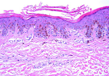 Fig. 1.2Junctional melanocytic nevus demonstrates small nests of melanocytes that can be differentiated from keratinocytes based upon morphologic features. Original magnification 200
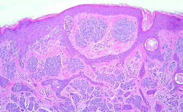 Fig. 1.3Compound melanocytic nevus has nests of nevus cells within the epidermis as well as within the dermis. Original magnification 100
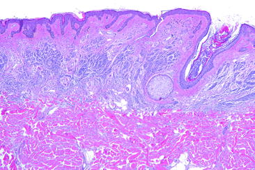
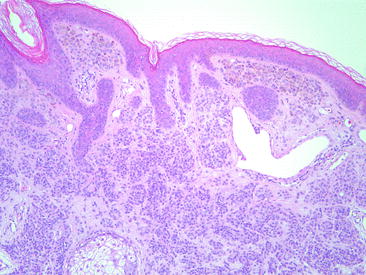 Fig. 1.4Intradermal nevus demonstrates nests of melanocytes restricted to the dermis with no epidermal involvement. Original magnification 100
- Acquired melanocytic nevus
- Histologic
- Junctional component should be almost entirely nested and sharply circumscribed
- Proliferation of single melanocytes is uncommon
- Pagetoid cells may occur secondary to trauma, in childhood, and in acral sites, but should not be abundant
- Pagetoid single or nested melanocytes located above the basal layer of the epidermis
- Presence implies loss of connection to basement membrane (through either trauma or deranged cellular substructure)
- Maturation in dermis (Fig. )
- Nevus cells become smaller and darker
- Abundant cytoplasm and vesicular nuclei in papillary dermis
- Minimal cytoplasm, small, dark nuclei at base
- Nests become smaller and eventuate in single melanocytes traversing between dermal collagen bundles
- Orderly maturation sequence is the rule absence raises possibility of melanoma
- Dermal mitoses rare should never be at base of lesion
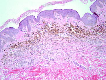 Fig. 1.5Maturation is a feature of benign melanocytic proliferations. The melanocytes become smaller and darker and the nests become smaller and more widely dispersed with progressive descent into the dermis. Pigmentation also tends to diminish with progressive descent. Original magnification 100
- Congenital melanocytic nevus
- Clinical
- Present in about 1% of newborns
- Often larger than acquired nevi
- May be hair-bearing
- So-called giant congenital nevi (>20 cm) often have a bathing suit distribution
- Incidence of developing melanoma
- Minimally increased in small congenital nevi
- May be as much as 10% in giant nevi
- Histologic (Figs. )
- Can be junctional, compound, or intradermal
- Abundant single melanocytes within epidermis in some congenital nevi in children
- Nevus nests extend into lower third of reticular dermis or into subcutis
- Nevus nests track down appendages
- Nevus nests often have a superficial perivascular dermatitis appearance at low magnification
- Scattered Pagetoid cells may be present in the central portion of congenital nevi, especially during the first year of life
- Pseudovascular spaces are often present and are due to dyscohesion of melanocytes within dermal nests (Fig. )
- Neurotization is commonly seen and is believed to be part of the maturation process (Fig. )
Font size:
Interval:
Bookmark:
Similar books «Epidermal Cell Tumors: The Basics»
Look at similar books to Epidermal Cell Tumors: The Basics. We have selected literature similar in name and meaning in the hope of providing readers with more options to find new, interesting, not yet read works.
Discussion, reviews of the book Epidermal Cell Tumors: The Basics and just readers' own opinions. Leave your comments, write what you think about the work, its meaning or the main characters. Specify what exactly you liked and what you didn't like, and why you think so.