Bruce R. Smoller - Dermatopathology: The Basics
Here you can read online Bruce R. Smoller - Dermatopathology: The Basics full text of the book (entire story) in english for free. Download pdf and epub, get meaning, cover and reviews about this ebook. year: 2009, publisher: Springer, genre: Home and family. Description of the work, (preface) as well as reviews are available. Best literature library LitArk.com created for fans of good reading and offers a wide selection of genres:
Romance novel
Science fiction
Adventure
Detective
Science
History
Home and family
Prose
Art
Politics
Computer
Non-fiction
Religion
Business
Children
Humor
Choose a favorite category and find really read worthwhile books. Enjoy immersion in the world of imagination, feel the emotions of the characters or learn something new for yourself, make an fascinating discovery.
- Book:Dermatopathology: The Basics
- Author:
- Publisher:Springer
- Genre:
- Year:2009
- Rating:3 / 5
- Favourites:Add to favourites
- Your mark:
Dermatopathology: The Basics: summary, description and annotation
We offer to read an annotation, description, summary or preface (depends on what the author of the book "Dermatopathology: The Basics" wrote himself). If you haven't found the necessary information about the book — write in the comments, we will try to find it.
Bruce R. Smoller: author's other books
Who wrote Dermatopathology: The Basics? Find out the surname, the name of the author of the book and a list of all author's works by series.


Dermatopathology: The Basics — read online for free the complete book (whole text) full work
Below is the text of the book, divided by pages. System saving the place of the last page read, allows you to conveniently read the book "Dermatopathology: The Basics" online for free, without having to search again every time where you left off. Put a bookmark, and you can go to the page where you finished reading at any time.
Font size:
Interval:
Bookmark:
- Epidermis varies in thickness with site, but ranges from < 0.1 mm on the eyelids to about 1 mm on acral sites
- Dermis varies in thickness with site, but ranges from 1 mm on the face to 4 mm (approximately) on the back
- Subcutaneous fat varies extensively in thickness (up to multiple cm)
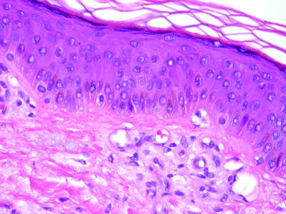
- Epidermis with stratified layers
- Stratum corneum
- Stratum granulosum
- Stratum spinosum
- Stratum basalis
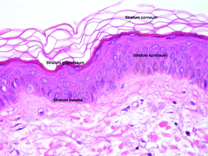
- Normal pattern basket-weave orthokeratosis
- Caused by increased lipid concentration in keratinocyte cytoplasm (Odland bodies)
- Basket-weave pattern is not seen on frozen section
- Nuclei are normally extruded before keratinocytes reach the stratum corneum
- Retained in conditions of dysmaturation and hyperproliferation (valuable clue to diagnoses)
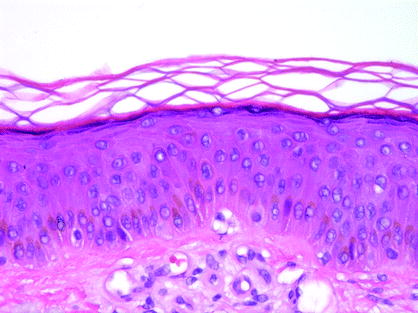
- Keratohyaline granules characterize this layer
- Profilaggrin involved in appropriate clumping of keratin in preparation for transformation to stratum corneum
- Histidine-rich granules
- Defect in profilaggrin seen in ichthyosis vulgaris (no granular layer present)
- Involucrin and keratolinin also appear as substrate for transglutaminase in cross linking of cellular envelope at this level
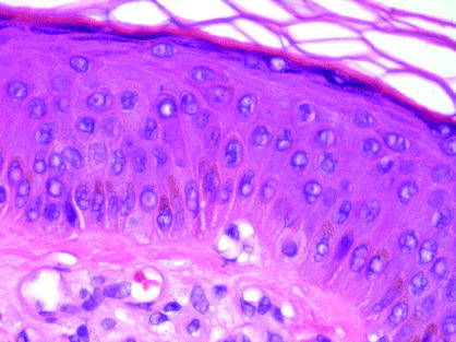
- Zone of maturation
- Takes basal cell 14 days to reach the stratum corneum and another 14 to desquamate under normal conditions
- Nuclear:cytoplasmic ratio becomes progressively smaller in these layers
- Keratin production switches from lower molecular weight (cytokeratin 5 and 14) to higher molecular weights (mainly keratins 1 and 10)
- Cells at base of rete ridges are stem cells that proliferate most rapidly
- Basal cells at tips of dermal papillae proliferate slowly
- Basal cells produce lowest molecular weight keratins (of all keratinocytes) keratins 5 and 14
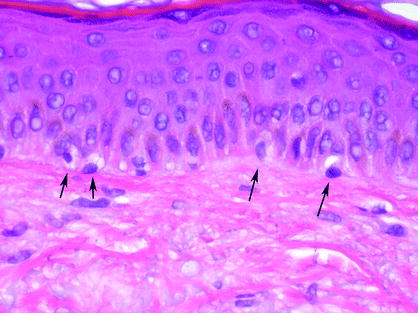
- Every tenth cell along basal layer is a melanocyte (on average)
- Increased in hyperpigmented and sun-exposed body sites
- Decreased on palms and soles
- Each melanocyte serves, via dendritic processes, 36 keratinocytes with melanin (on average)
- Increased numbers of melanocytes with sun exposure
- Produce eumelanin (brown and black) and phaeomelanin (red brown higher in sulfur content)
- Transfer melanosomes to lysosomes within keratinocytes via phagocytosis
- Individual variation in skin color is a function of relative numbers of stage IIV melanosomes. In general, all individuals have the same numbers of melanocytes (more stage IV in darker skin, more stage I in lighter skin)
- Darker races have larger, singly dispersed melanosomes in keratinocytes
- Caucasians have melanosome complexes within keratinocytes
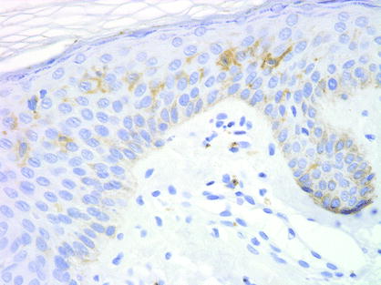
- Mid-epidermal dendritic cells
- Constitute 34% of all epidermal cells
- Involved in antigen presentation
- Bone marrow derived
- Express HLA-DR, ATPase, S100, CD1a
- Electron microscope demonstrates Birbeck granules, a pentalaminar structure shaped like a tennis racquet when viewed in full longitudinal section
- Increased numbers in conditions with increased antigen presentation (i.e., contact dermatitis)
- May coalesce into microgranulomata simulating nests of melanocytes

- Part of the sensory nervous system
- Sit on basement membrane
- Have desmosomal contacts with keratinocytes
- Haarscheibe one free nerve ending from dermis touches up to 50 Merkel cells to form this unit
Font size:
Interval:
Bookmark:
Similar books «Dermatopathology: The Basics»
Look at similar books to Dermatopathology: The Basics. We have selected literature similar in name and meaning in the hope of providing readers with more options to find new, interesting, not yet read works.
Discussion, reviews of the book Dermatopathology: The Basics and just readers' own opinions. Leave your comments, write what you think about the work, its meaning or the main characters. Specify what exactly you liked and what you didn't like, and why you think so.