• Complain

Haybaeck - Mechanisms of Molecular Carcinogenesis – Volume 2

Here you can read online Haybaeck - Mechanisms of Molecular Carcinogenesis – Volume 2 full text of the book (entire story) in english for free. Download pdf and epub, get meaning, cover and reviews about this ebook. City: Cham, year: 2017;2018, publisher: Springer International Publishing, genre: Romance novel. Description of the work, (preface) as well as reviews are available. Best literature library LitArk.com created for fans of good reading and offers a wide selection of genres:

Romance novel Science fiction Adventure Detective Science History Home and family Prose Art Politics Computer Non-fiction Religion Business Children Humor

Choose a favorite category and find really read worthwhile books. Enjoy immersion in the world of imagination, feel the emotions of the characters or learn something new for yourself, make an fascinating discovery.

Haybaeck Mechanisms of Molecular Carcinogenesis – Volume 2
  • Book:
    Mechanisms of Molecular Carcinogenesis – Volume 2
  • Author:
  • Publisher:
    Springer International Publishing
  • Genre:
  • Year:
    2017;2018
  • City:
    Cham
  • Rating:
    3 / 5
  • Favourites:
    Add to favourites
  • Your mark:
    • 60
    • 1
    • 2
    • 3
    • 4
    • 5

Mechanisms of Molecular Carcinogenesis – Volume 2: summary, description and annotation

We offer to read an annotation, description, summary or preface (depends on what the author of the book "Mechanisms of Molecular Carcinogenesis – Volume 2" wrote himself). If you haven't found the necessary information about the book — write in the comments, we will try to find it.

Haybaeck: author's other books


Who wrote Mechanisms of Molecular Carcinogenesis – Volume 2? Find out the surname, the name of the author of the book and a list of all author's works by series.

Mechanisms of Molecular Carcinogenesis – Volume 2 — read online for free the complete book (whole text) full work

Below is the text of the book, divided by pages. System saving the place of the last page read, allows you to conveniently read the book "Mechanisms of Molecular Carcinogenesis – Volume 2" online for free, without having to search again every time where you left off. Put a bookmark, and you can go to the page where you finished reading at any time.

Light

Font size:

Reset

Interval:

Bookmark:

Make
Springer International Publishing AG 2017
Johannes Haybaeck (ed.) Mechanisms of Molecular Carcinogenesis Volume 2 10.1007/978-3-319-53661-3_1
1. Molecular Classification of Breast Cancer
Iva Bri 1
(1)
Institute of Pathology, Medical University of Graz, Auenbruggerplatz 25, 8036 Graz, Austria
(2)
Division of Oncology, Department of Internal Medicine, Medical University of Graz, Auenbruggerplatz 15, 8036 Graz, Austria
Iva Bri (Corresponding author)
Email:
Marija Bali
Email:
1.1
1.2
1.2.1
1.2.2
1.3
1.3.1
1.3.2
1.3.3
1.3.4
1.3.5
1.4
1.4.1
1.5
Abstract
Breast cancer is the most common cancer in women, and it is one of the most intensively studied cancers. Out of all diagnosed breast cancer in women, only a small proportion develops in a familial setting, and for the large majority of women, the risk of developing breast cancer is less known. In this chapter, we describe in situ and invasive breast cancers in detail, referring to different immunohistochemical and molecular events, as well as the molecular classification of this heterogeneous disease. We address most common and well-established biomarkers currently performed in breast cancer diagnostics. Finally, in the last part, we describe the implications of current knowledge on the further directions and address possible future perspectives in breast carcinogenesis.
1.1 Introduction
Breast cancer (BC) is the most common cancer in women, accounting for more than 1,500,000 cases worldwide annually. In 2012, the incidence rate was 1,676,000 []. BC constitutes a heterogeneous group of different tumors characterized by variable clinical and morphological features with distinct molecular alterations/aberrations. The traditional classification of BC is based upon the assessment of histological type and grade. Patients age, tumor size, and lymph node status are also included in risk stratification and clinical treatment decisions. Immunohistochemical (IHC) analysis of receptor status on routinely processed tissue samples, namely, (1) expression of estrogen receptor (ER), (2) progesterone receptor (PR), and (3) overexpression/amplification of the human epidermal growth factor receptor 2 (HER2), also termed erythroblastosis oncogene B2 (ERBB2), is an imperative for complete pathological assessment, and their expression is the most important determinant of systemic treatment. The scoring of hormone receptors gives a better insight into potential response to endocrine treatment, and treatment with the humanized monoclonal antibody trastuzumab is largely dependent on the presence of HER2 either by immunohistochemistry or by in situ hybridization methods. Nonetheless, because of the genetic heterogeneity of breast cancers, histologically similar tumors may show different behaviors and responses to systemic therapies.
In the last two decades, different techniques have been used to collect molecular data on BC. By means of microarray-based gene expression profiling, the concept of heterogeneity among BCs has been established, demonstrating the need for distinct therapeutic approaches even further. More recently, next-generation sequencing (NGS) has brought new insights into BC classification. Prognostic and predictive subgroups have been suggested in order to enable individualized therapy [].
Here, we review histological features of ductal carcinoma in situ (DCIS) and histological subtypes of invasive BCs and current state of their molecular classifications and discuss the biological processes that are likely to play a role in BC carcinogenesis. Special subtypes and familial BCs will also be discussed, as well as future perspectives.
1.2 Histopathological Classification of In Situ and Invasive Breast Cancer
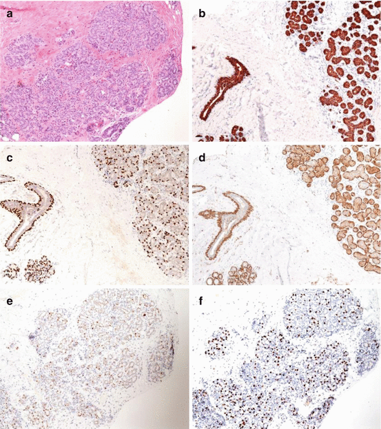
The human breast is composed of a branching duct system of 1520 lobes separated by fatty tissue. The terminal duct lobular units (TDLU) are the functional units of the breast and consist of the intralobular duct, ductules, and lobules that are lined by a single layer of cuboidal epithelium (luminal epithelium) supported by underlying basal/myoepithelial cells. The IHC profile of these cells differs: luminal epithelial cells can be highlighted by low molecular weight keratins, such as CK8, 18, and 19, whereas basal/myoepithelial cells stain with antibodies against p63, smooth muscle actin (SMA), smooth muscle myosin, calponin, S100-protein, CD10, CK5/6, etc. (Fig. ].
Fig 11 Normal breast tissue The terminal duct lobular unit TDLU is - photo 1
Fig. 1.1
Normal breast tissue. The terminal duct lobular unit (TDLU) is composed of the intralobular duct, ductules, and lobules lined by a single layer of luminal epithelium resting on a basal/myoepithelial layer ( a ). Luminal epithelium immunohistochemically highlighted by CK 8/18 ( b ). Basal layer immunohistochemically highlighted by p63 as nuclear staining ( c ) and SMA as cytoplasmic staining ( d ). Immunohistochemical expression of ER ( e ) and PR ( f ) seen as brown nuclear staining
1.2.1 Histological Features of Ductal Carcinoma In Situ
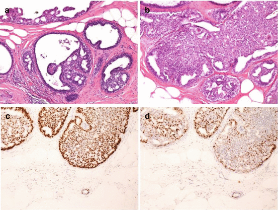
Ductal carcinoma in situ (DCIS) is characterized by proliferation of neoplastic luminal epithelial cells lined by a layer of myoepithelial cells and surrounded by an intact basement membrane, confined to the mammary ductal-lobular system. According to nuclear grade, differentiation, and presence of necrosis, they are classified into three grades: low, intermediate, and high grade [). The nuclei are uniform in size and shape and have regular chromatin. Microcalcifications can be found, but mitosis and necrosis are uncommon. Intermediate-grade DCIS is characterized by mild to moderately enlarged cells; nuclei have variably coarse chromatin. Microcalcifications, punctuate or comedo necrosis, and mitosis can be found. High-grade DCIS is composed of large atypical cells growing in solid, cribriform, or micropapillary pattern. Nuclei are pleomorphic, with coarse chromatin and prominent nucleoli. Mitotic figures, comedo necrosis with necrotic debris in lumina of the ducts, and amorphous microcalcifications are frequently seen.
Fig 12 Low-grade ductal carcinoma in situ Cribriform a and solid pattern - photo 2
Fig. 1.2
Low-grade ductal carcinoma in situ. Cribriform ( a ) and solid pattern ( b ) of growth characterized by small, uniform cells. Immunohistochemical expression of ER ( c ) and PR ( d ) seen as brown nuclear staining
1.2.2 Histological Types and Grade of Invasive Breast Cancers
The World Health Organization (WHO) classification recognizes ten main histological types of invasive BC with their subtypes and various rare entities (Table ]. Tumor cells can have abundant or eosinophilic cytoplasm; nuclei may be uniform or pleomorphic with prominent nucleoli. Mitotic figures can be absent or numerous. Necrosis may be present or absent, as well as DCIS.
Table 1.1
WHO classification of invasive breast carcinomas, without papillary lesions and epithelial-myoepithelial tumors []
Invasive breast carcinoma (types)
Classification
Invasive carcinoma of no special type (NST)
8500/3
Pleomorphic carcinoma
Next page
Light

Font size:

Reset

Interval:

Bookmark:

Make

Similar books «Mechanisms of Molecular Carcinogenesis – Volume 2»

Look at similar books to Mechanisms of Molecular Carcinogenesis – Volume 2. We have selected literature similar in name and meaning in the hope of providing readers with more options to find new, interesting, not yet read works.


Reviews about «Mechanisms of Molecular Carcinogenesis – Volume 2»

Discussion, reviews of the book Mechanisms of Molecular Carcinogenesis – Volume 2 and just readers' own opinions. Leave your comments, write what you think about the work, its meaning or the main characters. Specify what exactly you liked and what you didn't like, and why you think so.